Local mechanical properties in articular cartilage and other biological tissues are typically measured by tracking the displacement of cells, cell nuclei and other fiducial markers using particle image velocimetry (PIV) and other feature-tracking methods (see "Quasi-Static Shear Mechanical Properties of Articular Cartilage"). However, these techniques are limited in spatial resolution by the density of trackable markers. In adult articular cartilage, intervertebral disk and other soft tissues, cells can be very sparse. Therefore, we have developed grid-resolution automated tissue elastography (GRATE) and window averaged noisy differentiation (WAND), two techniques that allow for measurement of the local shear modulus with improved spatial resolution.
In GRATE, a grid of lines is photobleached onto images of fluorescently-stained biological tissue using a high-intensity laser. Below is a movie taken near the surface of a sample of articular cartilage with multiple vertical photbleached lines under sinusoidal shear. The top of the fluorescently stained (green) sample is the articular surface.
GRATE Movie from Cohen Group on Vimeo.
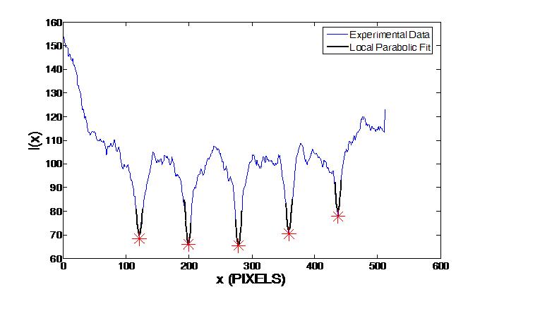
To determine local tissue deformation, the intensity I of each image at depth z from the surface as a function of horizontal location x is plotted and the local minima corresponding to each photobleached line is tracked as a function of depth from the surface and time.
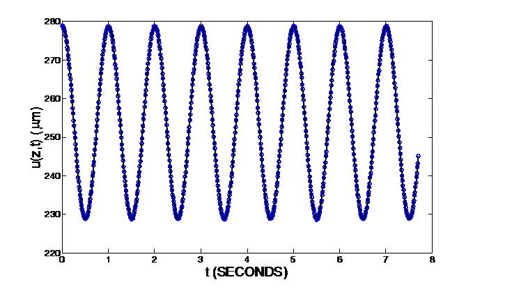
Below is a plot of the average photobleached line location at a depth z as a function of time. The solid line is a sinusoidal fit from which the amplitude and phase of u(z,t), the tissue displacement at a depth z, are determined.
Determination of the depth-dependent shear modulus profile of articular cartilage requires taking the derivative of often-noisy displacement data. To perform this differentiation, we use Window-Averaged Noisy Differentiation (WAND), a novel numerical technique developed by Attila Bergou (Cornell University). In WAND, the experimental error in the calculated derivative is minimized subject to the constraint that the truncation error (associated with the derivative approximation) remains small.
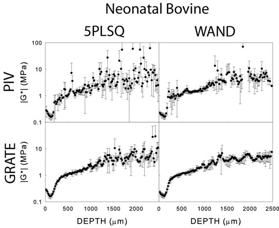
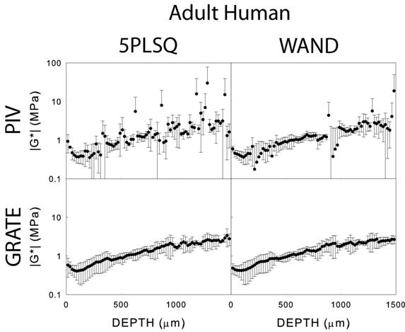
Below is a comparison of average shear modulus profiles for three samples of neonatal bovine and adult human articular cartilage sheared and analyzed at a resolution of 40 μm using PIV and GRATE with and without WAND. Data analyzed without WAND was differentiated using 5 point least-squares fitting (5PLSQ). Note that data analyzed using GRATE and WAND was more accurate, yielding profiles with smaller standard deviations.
For more information, see our paper (LINK TO PAPER).